Think It’s the Spine? It Might Be the SI Joint—Identify the Driver Before Imaging or Referral.

Cut through diagnostic uncertainty and confidently identify the true pain generator in complex low back cases. Patients feel heard when you bring clarity to complex pain—strengthening trust and guides better outcomes.

Who is he?

Diagnose the Circuit, Not Just the Symptom

My light bulb analogy helps illustrate why we need to go beyond treating the immediate pain and examine the entire “circuit” – the interconnected system of nerves, muscles, and biomechanics.

Let me explain.

When a light bulb burns out, the immediate thought is: replace the bulb.
But what if that doesn’t work? Do you check the switch? The wiring? Or maybe the fuse box?

I view each patient through this lens
💡 Injury site → Light bulb
🔌 Nerve entrapment → Wiring
 🎚 Spinal column → Switch
Brain → Fuse box

Now, let’s apply this to something simple—a sprained ankle.

A true sprain means damaged tissue—so why can someone walk normally one day but limp the next? If the tissue is torn, pain should be consistent, right?

So what else could be happening?
🔹 A peripheral nerve entrapment causing the limp?
🔹 An L5 nerve root involvement affecting motor control?
🔹 A brain-related issue, like an early sign of Parkinson’s? (Rare, but possible.)

By focusing on the entire circuit, we can identify the root cause of the problem and develop a more effective, long-term treatment plan. This approach leads to lasting recovery and prevents recurring issues.

Do you approach injuries this way?

Gemini_Generated_Image_o81gglo81gglo81g-min

Understanding Spinal Health: What MRI Might Be Missing

Imaging often reveals “abnormalities” like hip labral tears and lumbar disc bulges that are normal age-related changes, not necessarily sources of dysfunction — our commitment to recovery should stay rooted in MVMT, not in MRI results. 

  • MRI findings in asymptomatic patients: Research by Brinjikji et al. (2015) shows significant spinal degeneration (e.g., disc bulges, herniations) often appears in patients with no symptoms.

  • Spinal degeneration does not always correlate with pain: Just because degeneration is visible on an MRI doesn’t mean it’s the source of the patient’s pain.

  • Identify the source of pain: Early, accurate diagnosis of movement dysfunction and compensations can guide more effective treatment and reduce unnecessary imaging or referrals.

  • Comprehensive MVMT assessments: We assess how a patient’s spine loads during MVMT to pinpoint the dysfunctional pattern in their pain—not just what’s seen in an MRI.

  • When symptoms regress or stall, MRI findings may still help determine whether a shift from conservative to non-conservative care is warranted—but they shouldn’t lead care from the start.
Untitled (13)-min

Asymptomatic MRI Findings

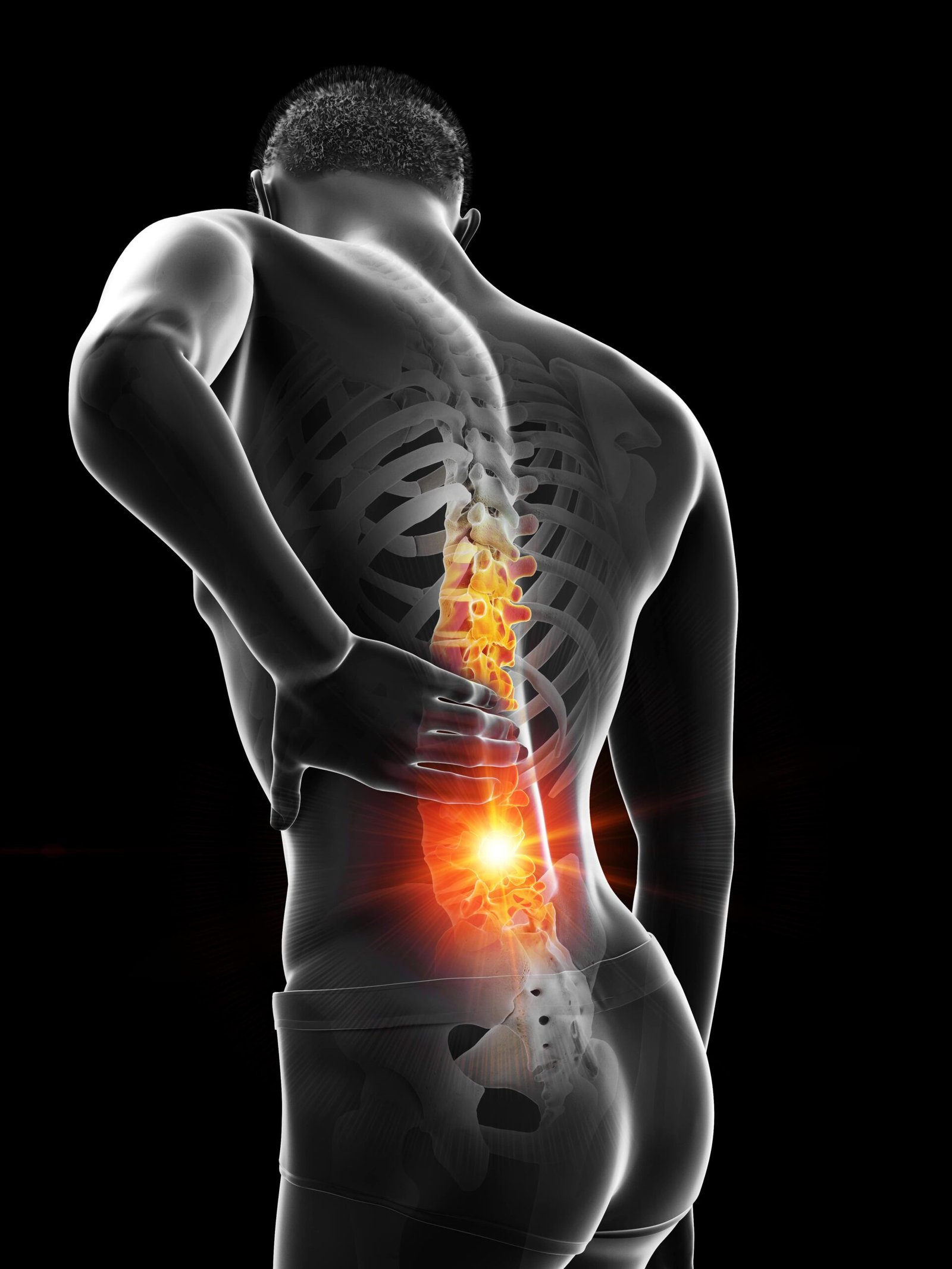
Degenerative Disc Diease
MRI studies show that 37% of 20-year-olds and 96% of 80-year-olds have signs of disc degeneration—most frequently seen in individuals in their 50s
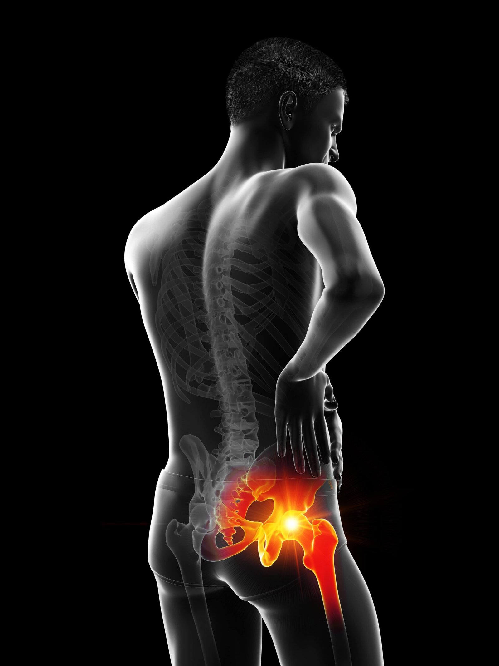
Hip Pathology
Individuals over 30 are 8.1x more likely to present with a labral tear. By age 35, they’re 13.7x more likely to show chondral defects.
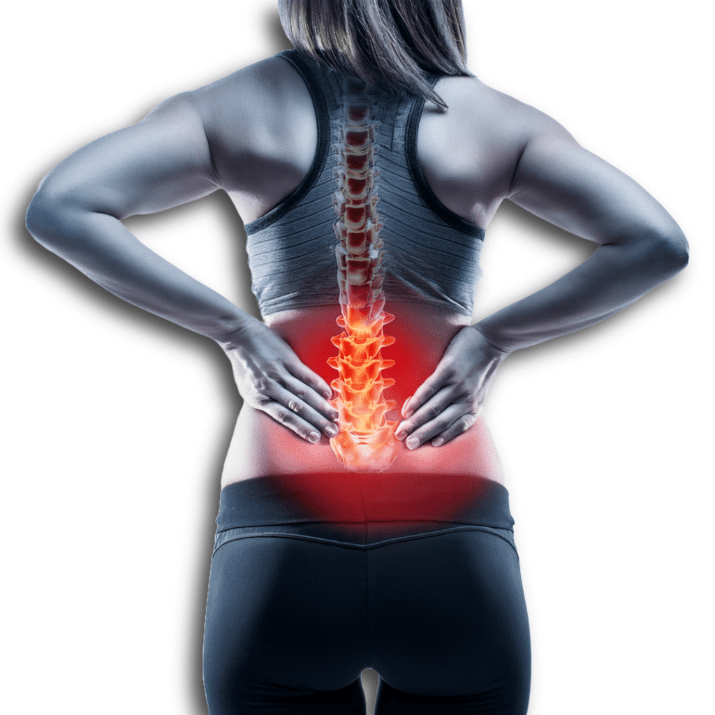
Disc Herniation
re present in 30% of 20-year-olds and 60% of 50-year-olds—even in the absence of symptoms.

Brinjikji et al. (2015) – MRI Findings of Disc Degeneration Are More Prevalent in Adults with Low Back Pain Than in Asymptomatic Controls: A Systematic Review and Meta-Analysis.

Register, B., Pennock, A. T., Ho, C. P., Strickland, C. D., Lawand, A., & Philippon, M. J. (2012). Prevalence of abnormal hip findings in asymptomatic participants: a prospective, blinded study. The American journal of sports medicine40(12), 2720-2724.

Header 1

What doctors say!

Nulla vitae elit libero, a pharetra augue. Morbi leo risus, porta ac consectetur ac, vestibulum at eros. Maecenas faucibus mollis interdum. Cum sociis natoque penatibus et magnis dis parturient montes, nascetur ridiculus mus. Praesent commodo cursus magna, vel scelerisque nisl consecteturet.

Dr. Bruce MT rowat

Internal Medicine

Clarify Low Back Pain vs SI joint in Under 5 Minutes—Using Clinical Reasoning, Not Just Imaging

Gain actionable insights to refine your exam process and confidently determine when to refer or take further action. Complete the form, and we'll set up a time that works best for you or your clinic.